Ultrasound scanners can be regarded as Medical Sonar, which uses ultrasonic waves to detect an object. Today, it is a highly familiar diagnostic technique. It’s current importance can be judged by the fact that, of all the various kinds of diagnostic images produced in the world, 1 in 4 is an ultrasound scan[1].
When did the story begin ?
Ultrasound has its roots lying back in history over 225+ years ago. Italian physiologist Lazzaro Spallanzani is often credited for discovering Ultrasonography. In 1794 Spallanzani performed sets of experiments on Bats and noted that bats can find the desired location based on the Echoes/reflected waves from its surrounding objects. Later discovery of Piezoelectricity in 1877 and invention of hydrophone/transducer in 1915 initiated further research. The use of ultrasound in medicine initially started with its application in therapy rather than diagnosis. However, many physician-scientists from different parts of Europe and America were working relentlessly to discover its diagnostic side. Finally in the late 1940s, George Ludwig at Naval Medical Research Institute, Bethesda, Maryland reported application of ultrasonic energy to the human body for diagnostic purposes[2]. Dr. Karl Theodore Dussik of Austria published the first paper on Medical Ultrasonics in 1942, he transmitted an ultrasound beam through the human skull in an attempt of detecting brain tumor[1]. In the 1950s Professor Ian Donald of Scotland developed practical technology and application for ultrasound[3]. And in 1963, this technology was finally available for general medical use. Since then Ultrasound imaging has come a long way.
So what is ultrasound and how does ultrasound imaging work?
Medical ultrasound lies within a frequency range of 30kHz to 500MHz, the lower frequency (30KHz to 3MHz) are for therapeutic purposes, the higher ones (2 to 40MHz) are for diagnosis and the very highest (50 to 500MHz) are for microscopic imaging[4]. Ultrasound diagnosis is based on two main techniques: The Pulse Echo Method and the Doppler Effect.
In Pulse echo method, a transducer produces sound waves which are then reflected back from organs and tissues. These reflected vibrations are then translated by ultrasound machine and transformed into an image using signal processing. Quality of the image is based on the depth and strength of the reflected vibrations/echos [3]. Conductive gels are used along with the transducers for better transmission of sound waves into the body. Pulse echo method can create an image of the structure inside the body but is not useful to trace moving entities.
Doppler effect is employed to measure the sound waves which are reflected by the moving objects such as red blood cells, the baseline is, doppler effect enables the anticipation of blood flow and muscle motion. In moving echogenic structures for example moving blood cells, echoes/reflections captured from the object travelling towards the transducer will have an increased frequency compared with the transmitted frequency, and echoes reflected off the object moving away from the transducer will have a lower frequency[4].
Ultrasound imaging is widely used in fields such as general medicine, internal medicine, gynaecology, obstetrics and veterinary medicine, all thanks to its numerous advantages like low maintenance and acquisition cost, no known side effects, risk free when used with right dosage, real time images, high spatial resolution.
Which factors triggered the further innovation/development?
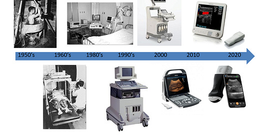
Like every other technology, Ultrasound also comes with its fair share of drawbacks. However, in order to understand the value of innovation it is important to be aware of how the technology was, when it made its debut. In the beginning, Ultrasound machines were too bulky and expensive for regular medical applications. In order to make it more user friendly and readily accessible as compared to other imaging modalities such as CT and MRI, focus was laid on the portability of the device. Ultrasound has undergone tremendous transition from conventional cart based system US to Pocket size hand held US and has shown significant development in terms of reduction in size, cost and at the same time improvement in computational power and portability. While considering portability of the device, Cable management was another topic of concern. Cables which connect transducers to the computing platform/cart were not only difficult to manage but also increased the risk of infection in sterile environments. In interventional suits where swift and accurate motions have a crucial role to play, managing cables in order to provide a wide scanning range and uninterrupted free movements at times can produce undesired hassles. And thus, the portable machines were born.
Portable ultrasound devices are available in three main types: Laptop associated devices, Hand carried and Hand held devices. According to recent trends, pocket sized handheld devices are gaining more popularity in the medical market. The Acuson P10 system by Siemens Medical Solutions was introduced in 2007 as the first portable ultrasound, it weighs about 0.73Kg and was designed for immediate and easy use in emergency medicine, cardiology and Obstetrics with an intuitive digital assistant interface[12]. In 2012, Siemens Healthcare introduced Acuson Freestyle wireless Ultrasound system. In this modality the communication between the console and transducers was established using Ultra-wideband (UWB) which worked 75 times faster than Bluetooth, it worked at a frequency of 8 gigahertz and could transmit 20 frames per seconds[5]. Acuson wireless ultrasound system allowed the user to operate a transducer upto 3m away from the system, which gave physicians a liberty to easily carry the scanner from one room to another. This pioneering breakthrough led many other healthcare companies to step up in this novel area of work. In 2012, GE healthcare brought the VScan Dual Probe in market, which is the first of its kind featured with two transducers in single probe. Dual probes facilitate deep and shallow scanning. Its design covers a broad spectrum of clinical applications such as ultrasound guided catheter placement, cardiac, abdominal, thoracic and foetal issues. As per the studies, VScan is practical, possibly aids diagnosis and prevents misdiagnosis [12,15].
To make the system even more compact and efficient, in 2015 a South Korea based company Healcerion came up with its first FDA approved wireless, app based ultrasound system[6]. This groundbreaking work laid the foundation of ultrasound transducers, compatible with most smartphones and tablets. At its core, these smartphone connected devices come with a software package for a smartphone. When coupled with a modified ultrasound transducer that plugs into the phone's USB port the software converts the phone into an ultrasound machine[7]. Currently the use of direct WiFi to connect to iOS and android apps is prevalent. Later in 2015 Phillips also launched its FDA cleared ultrasound device called Lumify. Smartphones tablet connected Lumify comes with three different transducers that extends its area of application including Cardiac, OB/GYN, Lung, Abdomen, FAST, Soft tissue, Vascular, Superficial and Musculoskeletal [12]. Lumify also provides tele-ultrasound solutions to remotely collaborate with clinicians and for training programs. Most of these wireless devices are compliant with cloud based data management system to store and consolidate the information.
In 2017, a startup Butterfly networks announced the first handheld ultrasound device featuring a silicon chip ( 2D array, 9000 micro machined sensor) instead of conventional piezoelectric crystal and began the era of Ultrasound on chip technology. In Ultrasound-on-chip technology, piezoelectric was replaced with micromachine CMUT (Capacitive micromachined ultrasound transducer) which would act like a tiny drum when voltage is applied in order to generate vibrations/ ultrasonic waves[16]. These ultrasound waves can emulate curved, linear or phased transducers at any time in M-, B Mode or Colour Doppler with 20-30 cm scan depth[12]. It can be connected to a smartphone that allows users to upload images to Butterfly cloud. This way, any expert with access can evaluate the ultrasound findings.
Generic timeline of Evolution
Benefits of the modern ultrasound system:
1. It provides a clear, real time view of the procedure with free movements, which is of high importance especially while performing delicate surgical procedures, proven feasible guiding tool for interventions [13,14]
2. With portable, cord free systems it is easy to maintain a sterile field which improves safety and reduces infection rate.
3. Improves imaging accessibility for the sites in the absence of reliable power sources for e.g. ambulance, remote areas, war zones etc.
4. Most of the portable ultrasound machines are Apple IOS + Android compatible, which makes it possible to share the scan instantly across multiple devices.
5. These compact devices can be charged by removable, rechargeable batteries which reduces the down time. Some can be also powered by paired devices.
6. Compact devices take less space in an emergency care unit loaded with multiple systems.
7. Cost effective at the same time enhances clinical efficiency through high quality data at the point of care.
8. Useful for training/ education purposes.
9. Due to its portability it is useful in pre-hospital settings. Provide help in rapid evaluation and triage of victims e.g. in context of mass casualty incident (MCI)[12]
More than comprehensive diagnosis, the main intention behind this portable technology is to provide quick answers to specific clinical questions.
Do they have any drawbacks ?
Although many studies have shown that there are no clinically relevant discrepancies in diagnosis performed with portable handheld ultrasounds. However, some have raised concerns regarding poor penetration of portable ultrasound scanners which limits the assessment of solid pathologies and deeper structures[9]. Another drawback is when most smartphones will have the capability to add an ultrasound probe at a low cost, the technology will be available to a wide layman audience with no training whatsoever which will drive the public image of ultrasound from one diagnostic machine to a means of producing unique entertainment images and videos[10]. Additional attention towards data transmission confidentiality and security of patient’s data is equally essential.
Portable ultrasound modality is definitely a technology jump with a significant impact. As with all of sonography, the result of an examination not only depends on the technology but also on the skill level of the operator therefore, a great deal of research and work remains to be done in order to achieve the desired outcomes[10]. A recent report looking at portable ultrasound devices predicts the market will grow 8.5 percent year over a year from 2018 to 2024. Adding to current technology, efforts are being taken by many companies to infuse Artificial Intelligence (AI) into the ultrasound system which will help operators to read scans and can also assist to predict correct diagnosis. One such example is Portland based startup YourLabs. Recently GE healthcare received FDA clearance for their Vivid Ulta Edition, an AI powered cardiovascular ultrasound system to shorten diagnosis time and improve measurement consistency. Ultrasound is a promising technology after overcoming the present shortcomings. Will it be possible to turn it into a future stethoscope? That remains to be seen...
References:
Woo J. Hong Kong (2002) “A history of the development of ultrasound in obstetrics and gynecology”, LINK
The history of ultrasound sonography, Ultrasound school info, LINK
Bellis, Mary (2021) “The history of ultrasound in Medicine”, LINK
Katherine A. Kaproth Joslin et.al.(2014) “The history of US: From bats and boats to the bedside and beyond”, LINK
“The first wireless ultrasound transducer”, Healthcare in Europe.com, LINK
“The world's first wireless app based ultrasound system interview with Dr. Benjamin Ryu, CEO of Healcerion”, LINK
Rob Goodier (2011) “Ultrasound is now on Smartphones for change” Engineering for change, LINK
Robinson T.M.(2007) “Basic principles of ultrasound physics for medical imaging applications” NATO Science series, Vol240. Springer, Dordrecht, LINK
Ka Hei Tse et.al.(2014) “Pocket sized versus standard ultrasound machines in abdominal imaging” Singapore Med J, Vol55(6), LINK
Delores Jones(2014) “Smartphones compatible ultrasound probe” Journal of diagnostic medical sonography”, Vol30(4), LINK
Tadashi Kobyashi (2016) “ Development of pocket size handheld ultrasound device enhancing peoples abilities and need on educating on them” Journal of general and family medicine”, Journal of general and family medicine, Vol17(4)276-288, LINK
Clevert DA et al. (2019) “ESR statement on portable ultrasound devices”, Insights imaging 10, 89, LINK
Keil-Rios D et al. (2016) “Pocket ultrasound device as a complement to physical examination for ascites evaluation and guided paracentesis”, Intern Emerg Med 11(3): 461-466, LINK
Michon A et. al.(2019) “Use of pocket sized ultrasound in internal medicine (hospitalist) practice: Feedback and perspectives”, Rev Med Intern 40(4):200-225
A Colclough et. al.(2017) “ Pocket sized point of care cardiac ultrasound devices”, Herz, Vol42:255-261, LINK
Eliza Strickland(2017) “New ultrasound on chip tool could revolutionize medical imaging” IEEE Spectrum LINK


This writing on “Evolution of Ultrasound Machines from Conventional to
Wireless Pocket Scanners” is and very informative / highly appreciated.
Specially ‘Generic timeline of Evolution’ shown by photographs is very nice
(photo summary in very brief yet informative).
Agreed that in this assay you have not discussed particular/specific
applications of ‘Ultrasound’ technology’, some mention of
‘The Role of Ultrasound in Breast Cancer Screening’ (compare to
‘Mammography’ which is the gold standard for breast cancer screening)
was expected [may not be odd/out-of-place, I guess. With increasing
awareness among patients (about inconvenience) and health care
providers of mammography limitations especially in dense breasts,
supplemental screening for breast cancer with ultrasound and magnetic
resonance imaging has been expanding. There are many articles/reviews
on the efficacy, utility, and feasibility of ultrasound as a screening tool
for the early detection of occult breast cancer these days on this well
debated topic of current importance {which may be referred}.
Without that also it is excellent [added just for information which is
Known to you, I am sure]. Go ahead. All the best.